When you want to find radiology books mri, you may need to consider between many choices. Finding the best radiology books mri is not an easy task. In this post, we create a very short list about top 12 the best radiology books mri for you. You can check detail product features, product specifications and also our voting for each product. Let’s start with following top 12 radiology books mri:
Reviews
1. Fundamentals of Body MRI, 2e (Fundamentals of Radiology)
Description
Effectively perform and interpret MR body imaging with this concise, highly illustrated resource! Fundamentals of Body MRI, 2nd Edition, by Drs. Christopher Roth and Sandeep Deshmukh, covers the essential concepts residents, fellows, and practitoners need to know, laying a solid foundation for understanding the basics and making accurate diagnoses. This easy-to-use title in the Fundamentals of Radiology series covers all common body MR imaging indications and conditions, while providing new content on physics and noninterpretive skills with an emphasis on quality and safety.
- More than 1,400 detailed MRI images and 100 algorithms and diagrams highlight key findings and help you grasp visual nuances of images youre likely to encounter.
- All common body MR imaging content is covered, along with discussion of how physics, techniques, hardware, and artifacts affect results.
- Expert Consult eBook version included with purchase. This enhanced eBook experience allows you to search all of the text, figures, and references from the book on a variety of devices.
- Newly streamlined format helps you retrieve important information more quickly.
- Extensively revised content on the liver, including new MRI contrast agents; new coverage of the spleen; and new safety tips and guidelines keep you up to date.
- New chapters on GI imaging, the prostate, and the male genitourinary system make this a one-stop reference to address the full range of body MRI.
2. MRI of the Upper Extremity: Shoulder, Elbow, Wrist and Hand
Description
MRI of the Upper Extremity is a complete guide to MRI evaluation of shoulder, elbow, wrist, hand, and finger disorders. This highly illustrated text/atlas presents a practical approach to MRI interpretation, emphasizing the clinical correlations of imaging findings. More than 1,100 MRI scans show normal anatomy and pathologic findings, and a full-color cadaveric atlas familiarizes readers with anatomic structures seen on MR images.
Coverage of each joint begins with a review of MRI anatomy with cadaveric correlation and proceeds to technical MR imaging considerations and clinical assessment. Subsequent chapters thoroughly describe and illustrate MRI findings for specific disorders, including rotator cuff disease, nerve entrapment syndromes, osteochondral bodies, and triangular fibrocartilage disorders.
3. PET-CT and PET-MRI in Oncology: A Practical Guide (Medical Radiology)
Feature
Used Book in Good ConditionDescription
Over the past decade, PET-CT has achieved great success owing to its ability to simultaneously image structure and function, and show how the two are related. More recently, PET-MRI has also been developed, and it represents an exciting novel option that promises to have applications in oncology as well as neurology. The first part of this book discusses the basics of these dual-modality techniques, including the scanners themselves, radiotracers, scan performance, quantitation, and scan interpretation. As a result, the reader will learn how to perform the techniques to maximum benefit. The second part of the book then presents in detail the PET-CT and PET-MRI findings in cancers of the different body systems. The final two chapters address the use of PET/CT in radiotherapy planning and examine areas of controversy. The authors are world-renowned experts from North America, Europe, and Australia, and the lucid text is complemented by numerous high-quality illustrations.
4. Magnetic Resonance Imaging in Orthopaedics and Sports Medicine (2 Volume Set)
Description
Now in two volumes, the Third Edition of this standard-setting work is a state-of-the-art pictorial reference on orthopaedic magnetic resonance imaging. It combines 9,750 images and full-color illustrations, including gross anatomic dissections, line art, arthroscopic photographs, and three-dimensional imaging techniques and final renderings. Many MR images have been replaced in the Third Edition, and have even greater clarity, contrast, and precision.
5. MRI: Basic Principles and Applications
Description
This fifth edition of the most accessible introduction to MRI principles and applications from renowned teachers in the field provides an understandable yet comprehensive update.- Accessible introductory guide from renowned teachers in the field
- Provides a concise yet thorough introduction for MRI focusing on fundamental physics, pulse sequences, and clinical applications without presenting advanced math
- Takes a practical approach, including up-to-date protocols, and supports technical concepts with thorough explanations and illustrations
- Highlights sections that are directly relevant to radiology board exams
- Presents new information on the latest scan techniques and applications including 3 Tesla whole body scanners, safety issues, and the nephrotoxic effects of gadolinium-based contrast media
6. Magnetic Resonance Imaging of the Brain and Spine (2 Volume Set)
Feature
Used Book in Good ConditionDescription
Established as the leading textbook on imaging diagnosis of brain and spine disorders, Magnetic Resonance Imaging of the Brain and Spine is now in its Fourth Edition. This thoroughly updated two-volume reference delivers cutting-edge information on nearly every aspect of clinical neuroradiology. Expert neuroradiologists, innovative renowned MRI physicists, and experienced leading clinical neurospecialists from all over the world show how to generate state-of-the-art images and define diagnoses from crucial clinical/pathologic MR imaging correlations for neurologic, neurosurgical, and psychiatric diseases spanning fetal CNS anomalies to disorders of the aging brain. Highlights of this edition include over 6,800 images of remarkable quality, more color images, and new information using advanced techniques, including perfusion and diffusion MRI and functional MRI.
A companion Website will offer the fully searchable text and an image bank.
7. Textbook of Diagnostic Sonography: 2-Volume Set, 7e (Textbook of Diagnostic Ultrasonography)
Description
Stay up to date with the rapidly changing field of medical sonography! Heavily illustrated and extensively updated to reflect the latest developments in the field, Textbook of Diagnostic Sonography, 7th Edition equips you with an in-depth understanding of general/abdominal and obstetric/gynecologic sonography, the two primary divisions of sonography, as well as vascular sonography and echocardiography. Each chapter includes patient history, normal anatomy (including cross-sectional anatomy), ultrasound techniques, pathology, and related laboratory findings, giving you comprehensive insight drawn from the most current, complete information available.
- Full-color presentation enhances your learning experience with vibrantly detailed images.
- Pathology tables give you quick access to clinical findings, laboratory findings, sonography findings, and differential considerations.
- Sonographic Findings highlight key clinical information.
- Key terms and chapter objectives help you study more efficiently.
- Review questions on a companion Evolve website reinforce your understanding of essential concepts.
- New chapters detail the latest clinically relevant content in the areas of:
- Essentials of Patient Care for the Sonographer
- Artifacts in Image Acquisition
- Understanding Other Imaging Modalities
- Ergonomics and Musculoskeletal Issues in Sonography
- 3D and 4D Evaluation of Fetal Anomalies
- More than 700 new images (350 in color) clarify complex anatomic concepts.
- Extensive content updates reflect important changes in urinary, liver, musculoskeletal, breast, cerebrovascular, gynecological, and obstetric sonography.
8. Magnetic Resonance Imaging Handbook
Description
Magnetic resonance imaging (MRI) is a technique used in biomedical imaging and radiology to visualize internal structures of the body. Because MRI provides excellent contrast between different soft tissues, the technique is especially useful for diagnostic imaging of the brain, muscles, and heart.
In the past 20 years, MRI technology has improved significantly with the introduction of systems up to 7 Tesla (7 T) and with the development of numerous post-processing algorithms such as diffusion tensor imaging (DTI), functional MRI (fMRI), and spectroscopic imaging. From these developments, the diagnostic potentialities of MRI have improved impressively with an exceptional spatial resolution and the possibility of analyzing the morphology and function of several kinds of pathology.
Given these exciting developments, the Magnetic Resonance Imaging Handbook is a timely addition to the growing body of literature in the field. Comprised of three volumes, this comprehensive handbook:
- Covers the technology and practice of MRI from physical principles to cutting-edge applications
- Discusses MRI of the neck, brain, cardiovascular system, thorax, abdomen, pelvis, and musculoskeletal system
- Highlights each organs anatomy and pathological processes with high-quality images
- Examines the protocols and potentialities of advanced MRI scanners such as 7 T systems
- Includes extensive references at the end of each chapter to enhance further study
Thus, the Magnetic Resonance Imaging Handbook provides radiologists and imaging specialists with a valuable, state-of-the-art reference on MRI.
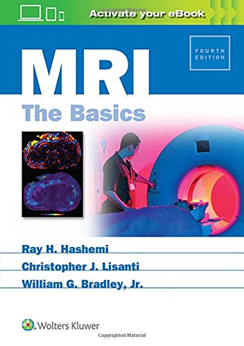
9. MRI: The Basics
Description
Feautures:
- Contains all-new chapters on general MR safety and contrast safety, as well as a new chapter on motion correction.
- Addresses timely topics such as susceptibility-weighted imaging (including other potential uses beyond hemorrhage detection), Restriction Spectrum Imaging (RSI), MR elastography, and MR relaxometry.
- Provides 100 new board-style questions in a separate chapter, as well as problem-solving and multiple choice questions in each chapter.
- Includes key points at the end of each chapter for quick reference and review.
- Ideal for radiologists, radiology residents and fellows, and radiologic technologists, as well as other professionals who encounter MRI in their practice, and those preparing for exams.
Your book purchase includes a complimentary download of the enhanced eBook for iOS, Android, PC & Mac. Take advantage of these practical features that will improve your eBook experience:
- The ability to download the eBook on multiple devices at one time providing a seamless reading experience online or offline
- Powerful search tools and smart navigation cross-links that allow you to search within this book, or across your entire library of VitalSource eBooks
- Multiple viewing options that enable you to scale images and text to any size without losing page clarity as well as responsive design
- The ability to highlight text and add notes with one click
10. Sectional Anatomy by MRI and CT, 4e
Feature
ElsevierDescription
Now available with state-of-the-art digital enhancements, the highly anticipated 4th edition of this classic reference is even more relevant and accessible for daily practice. A sure grasp of cross sectional anatomy is essential for accurate radiologic interpretation, and this atlas provides exactly the information needed in a practical, quick reference format. New color coding of anatomic structures and new scroll and zoom capabilities on photos in the eBook version make this title an essential diagnostic tool for both residents and practicing radiologists.
- Expert Consult eBook version included with purchase. This enhanced eBook experience allows you to search all of the text, figures, images, and references from the book on a variety of devices.
- Color-coded labels for nerves, vessels, muscles, bone tendons, and ligaments facilitate accurate identification of key anatomic structures.
- Scroll and zoom capabilities on photos in the accompanying eBook version enable easier accessibility during interpretation sessions and real-time resident education.
- Carefully labeled MRIs for all body parts, as well as schematic diagrams and concise statements, clarify correlations between bones and tissues.
- CT scans for selected body parts enhance anatomic visualization.
- More than 2,300 state-of-the-art images can be viewed in three standard planes: axial, coronal, and sagittal.
11. Breast MRI Teaching Atlas
Description
This atlas serves as a basic introduction to breast MRI. Organized by case, it emphasizes pertinent breast MRI findings and common indications for breast MRI. Topics include breast MRI basics, benign findings, breast malignancy, high-risk conditions, interesting cases, and breast implants. Brief teaching points accompany each case and highlight the importance of the findings. Breast MRI Teaching Atlas is an ideal resource for diagnostic radiologists, residents, fellows, and clinicians involved in the care of breast cancer patients, including surgeons, oncologists, and obstetricians/gynecologists.
12. Liver Imaging: MRI with CT Correlation (Current Clinical Imaging)
Description
- The first single source work to deal with the two primary radiologic modalities in diagnosing and treating benign and malignant diseases of the liver, presented with clearly laid out MRI and CT correlations. Developed by an editor team led by one of the worlds leading authorities in abdominal imaging, Richard C. Semelka MD.
- User-friendly, atlas-style presentation, with over 1500 MRI and CT images in over 320 figures featuring state-of-the-art MR and CT imaging sequences, multidetector row CT images, 3D reformatted images, breath-hold MRI sequences, and cutting-edge MR 3T images
- Highly practical approach for imaging of focal and diffuse liver lesions, complete relevant and systematic (differential) diagnostic information, the latest references to primary literature and clinical evidence, and patient management possibilities
- Reflects a pattern-recognition approach to MRI and CT imaging, assisting with efficient scanning of images and assessment and diagnosis of disorders


